[테크월드뉴스=조명의 기자]
The complex structure and thin thickness of the lung made it difficult to create an experimental artificial lung, but the POSTECH research team recently succeeded in making the artificial lung model by 3D printing.

POSTECH’s research team has succeeded in creating a three-dimensional lung model containing a variety of human alveolar cell lines using inkjet bioprinting. In particular, the inkjet bioprinting used in the research is attracting attention as it can produce and standardize patient-customized tissues, and can replace the existing test model as it can be mass produced.
The study was recently published in the international journal’Advanced Science’.
The human lungs constantly breathe in order to receive oxygen, which is necessary for vital activity, and to expel carbon dioxide, which is a by-product. Oxygen entering the body arrives at the alveoli through the airways, and is replaced with carbon dioxide carried by blood through the capillaries of the alveoli.
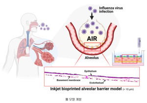
Here, the alveoli are made of a thin layer of epithelial cells, surrounded by thin capillaries around them, and have the shape of hollow grape clusters. The alveolar membrane, through which oxygen and carbon dioxide move, has a three-layer structure of’epithelial layer-basement membrane-endothelial capillary layer’ and has a very thin thickness for easy gas movement. Until now, there have been limitations in accurately replicating alveoli with such a thin and complex structure.
For the first time, the research team reproduced a three-layer alveolar barrier model with a thickness of about 10 μm by laminating alveolar cells at high resolution using drop-on-demand high-precision inkjet printing. The model thus produced showed a high degree of simulation when compared to a two-dimensional cell culture model as well as a three-dimensional unstructured model cultured by mixing alveolar cells and hydrogel.
In addition, the research team found that the produced alveolar barrier model similarly reproduced the physiological response at the actual tissue level in terms of viral infectivity and antiviral response. When this model was used as an influenza virus infection model, it was observed that virus self-proliferation and antiviral response appeared.
Professor Seongjoon Jeong said, “We print cells and fabricate tissues using bioprinting, but it is the first in the world to simulate the barrier of the alveoli, which has a three-layer structure of about 10 μm thickness.” It is also the first time that an antiviral reaction has been observed.”
In addition, “the artificial tissue produced this time can be used as an initial platform for evaluating the effectiveness of therapeutic drugs and vaccines capable of responding to infectious respiratory viruses, including novel coronavirus infections, as it enables mass production and quality control as well as patient-specific disease model production. Added.
On the other hand, this research was conducted with the support of the Korea Research Foundation’s mid-sized researcher support project, leading research center support project, and basic research project.
