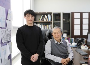
![(From left) Dr. Heejin Lim and DGIST Professor Daewon Moon. [사진=DGIST 제공]](https://i0.wp.com/cdn.hellodd.com/news/photo/202102/91674_303027_4938.jpg?w=560&ssl=1)
A technology that visualizes bio-imaging technology, which is a core technology necessary for early diagnosis of diseases or development of new drugs, is developed without distortion in an ultra-high vacuum environment. “Innovative technology for imaging living cells in ultra-high vacuum,” said Ron Heeren, a professor at Maastricht University in the Netherlands, a world-renowned mass spectrometry expert. “Interaction between drugs and cell membranes, catalytic surface research in solution, It is predicted that it will be used in mass spectrometry laboratories around the world for research on bio-living tissue chip devices.”
DGIST (President Kook Yang) is a mass spectrometric bio-imaging that visualizes the composition of living cell membrane molecules without distortion in an ultra-high vacuum environment by the research team of Dae-won Moon, Chair Professor of New Biology (concurrent position) and Hee-jin Lim and Ph.D. in New Biology. It announced on the 7th that it had developed the technology. It is expected to contribute greatly to elucidate complex disease mechanisms such as dementia and cancer.
Bio-imaging technology is a technology that makes it possible to see phenomena occurring in cells as images. Convergence of several fields such as biotechnology, physics, chemistry, and mechanical electronics is essential. For this reason, electron microscopy or SIMS (Secondary Ion Mass Spectrometry) analysis method using an accelerated electron beam or an accelerated ion beam in an ultra-high vacuum environment is mainly applied for advanced nano-imaging analysis. SIMS analysis is a technology mainly used for analyzing trace impurities for semiconductor manufacturing using accelerated ions, and its sensitivity is very high, so studies to apply it to bio-imaging technologies are active.
Currently, the cell analysis method using SIMS is a method in which living cells cultured in a solution are immobilized or cooled by a chemical method and then dried in an ultra-high vacuum environment. However, in this process, it was difficult to perform accurate analysis, such as distorting the unique molecular composition and distribution information of cells.
The research team has a 5 microliter (μl, 1 in 1 million liter) volume of micro-culture solution storage and a 1 micron (μm, 1 in 1 million liter) volume of cell culture solution under the substrate for culturing cells to enable analysis of cell membranes in a living state. Made thousands of metric) diameter holes. This was covered with a thin film of collagen biomolecules to facilitate cell adhesion and culture.
In a living state, the cultured cells were covered with monolayer graphene and introduced into an ultra-high vacuum environment. Single-layer graphene has a structure in which water molecules cannot leak out, and it is mechanically strong and can overcome water vapor pressure at room temperature. For this reason, the cells in the cell culture solution could be covered and maintained in an ultra-high vacuum environment. The research team was the first to succeed in imaging by applying the SIMS assay while protecting living cells.
The result of this study is to conduct convergence studies in various fields such as semiconductor engineering, nanomaterial engineering, biology, surface chemistry, theoretical chemistry, such as semiconductor process technology, graphene nanomaterial technology, cell culture, SIMS analysis technology, and one-dimensional dynamics theory calculation. It is significant in that it was performed through.
Professor Moon Dae-won said, “With the state-of-the-art nano-imaging technology, we are able to provide accurate mass spectrometric imaging of a variety of molecular information of living cell membranes without distortion.” “Understanding various phenomena such as corrosion, abrasion, catalysts, etc. occurring in liquid phases It will be a groundbreaking base technology.”
This research, which was conducted in collaboration with the research team of Professor Seo Dae-ha of DGIST Department of New Materials Science and the research team of professor Jang Yoon-hee of energy engineering, was published in’Nature Method (IF: 30.822)’ on the 4th.

Send SNS article
